SERVICES
ANIDOS provides a range of advanced animal tooth treatment, oral surgeries and medical therapies for cats and dogs.
Oral Medicine
The diagnosis and medical management of disease in the oral cavity and its supporting structures. The process usually starts with a consultation and examination of the awake animal. More information may be required from a detailed extra oral and intraoral examination which may involve periodontal charting, dental radiography, incisional biopsies and other diagnostic sample collection may be performed under general anaesthesia or sedation as indicated. In some cases advanced imaging modalities such as computed tomography (CT), and magnetic resonance imaging (MRI) may be required.
Typical cases include: feline chronic gingivostomatitis (FCGS), feline juvenile gingivitis, feline oral pain syndrome (FOPS), canine chronic ulcerative stomatitis (CCUS – formerly known as chronic ulcerative paradental stomatitis, CUPS), eosinophilic granulomas, masticatory muscle myositis (MMM), craniomandibular osteopathy (CMO), sialadenitis, necrotising sialometaplasia, necrotising osteomyelitis of the jaws, oral lesions due to electrical or chemical burns, oral masses, open-mouth jaw locking in cats, conservative management of jaw fractures with tape muzzles, the placement of oesophageal feeding tubes for nutrition, the non-surgical management of oronasal fistulas, etc.
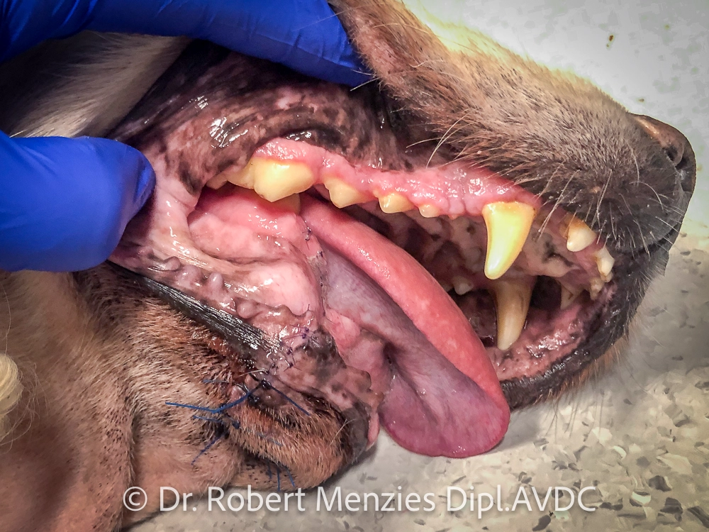
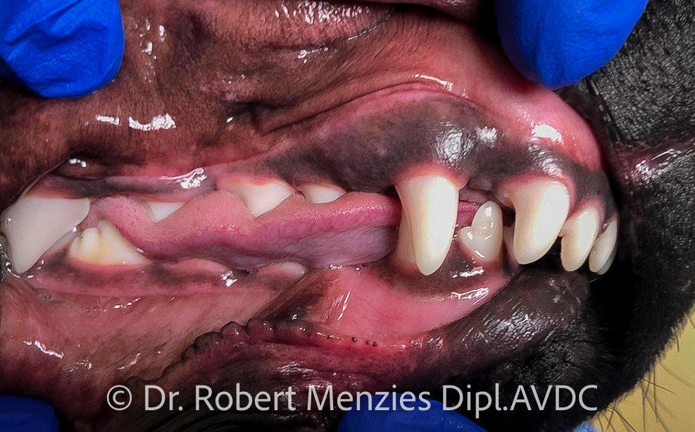
Sialadenitis
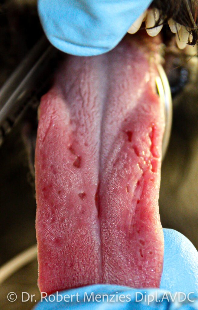
Palatitis & glossitis
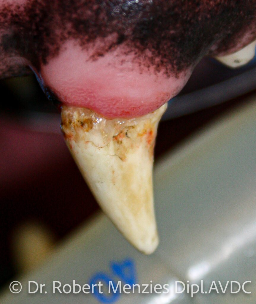
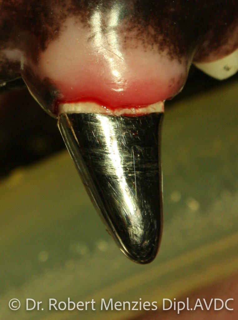
Endodontics
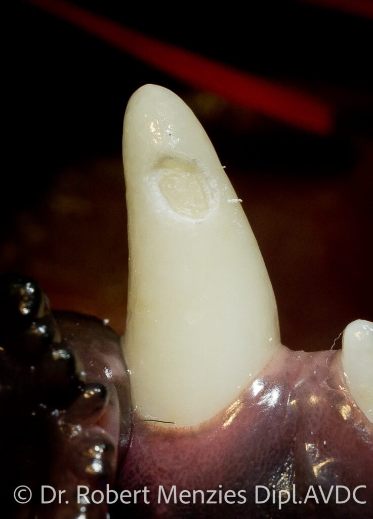
Therapies involving the pulp cavity with the aim of either preserving the tooth in its vital or non-vital state. Vital pulp therapy is aimed at keeping the tooth alive, such as a recently fractured tooth with pulp exposure (within 48hours for mature animals), or a crown reduction procedure to prevent a maloccluding lower canine tooth from traumatising the hard palate. Root canal therapy involves removing an irreversibly inflamed or necrotic pulp, disinfection of the pulp cavity, obturation and restoration of the access site.
The aim is to preserve the tooth despite it being non-vital. Indications include fractured teeth, splinted avulsed and luxated teeth, and infected teeth. The process may be done in one visit or it may require multiple visits. Endodontics therapies require ongoing monitoring for the life of the tooth.
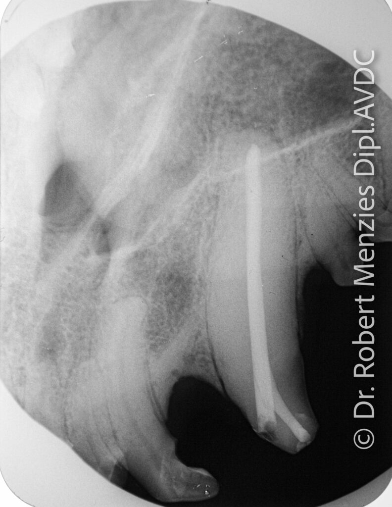
Root canal therapy
crown fracture of the left upper canine tooth in an 8-year-old Newfoundland dog.
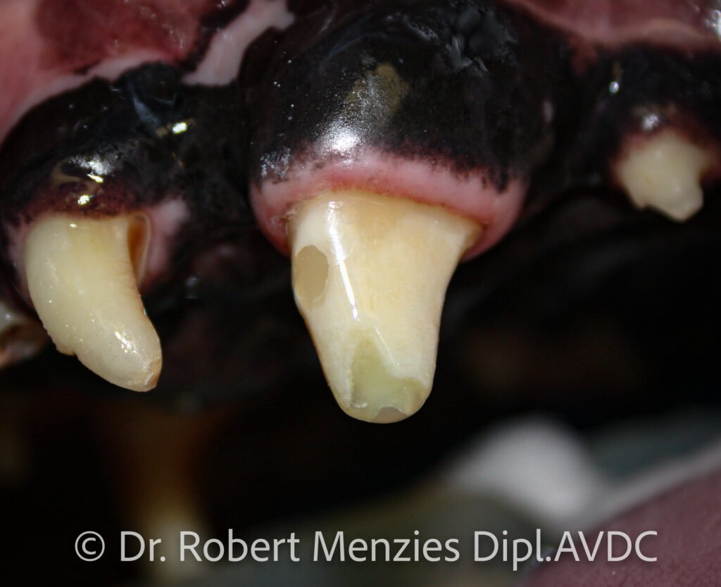
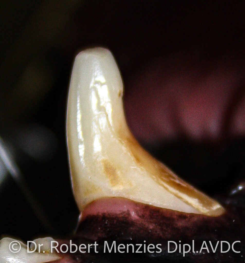
Vital pulp therapy
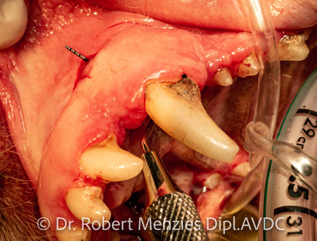
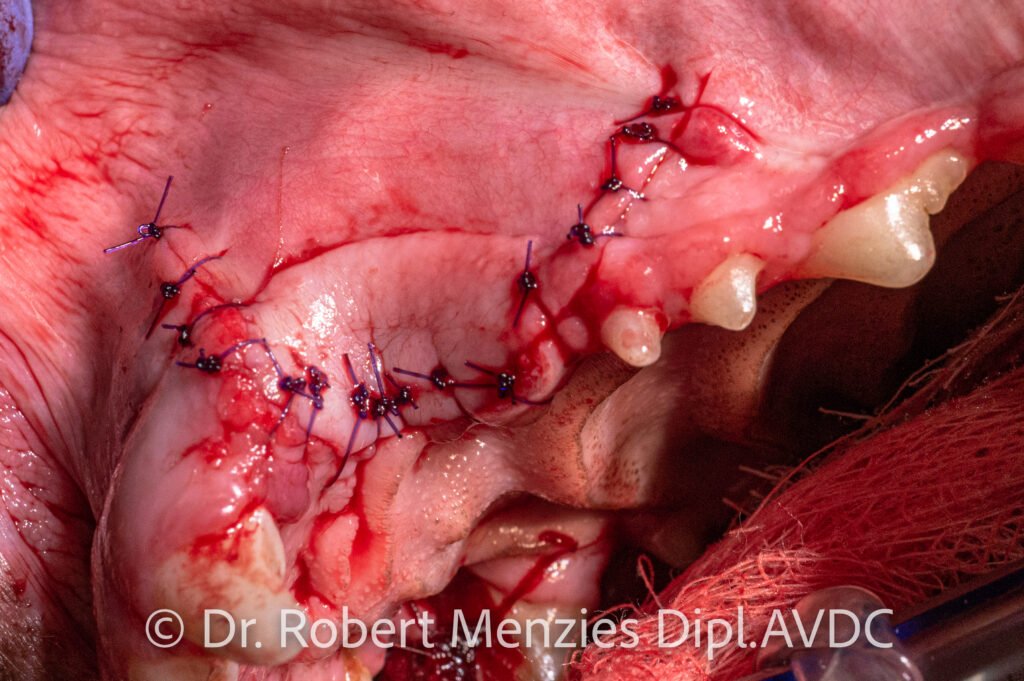
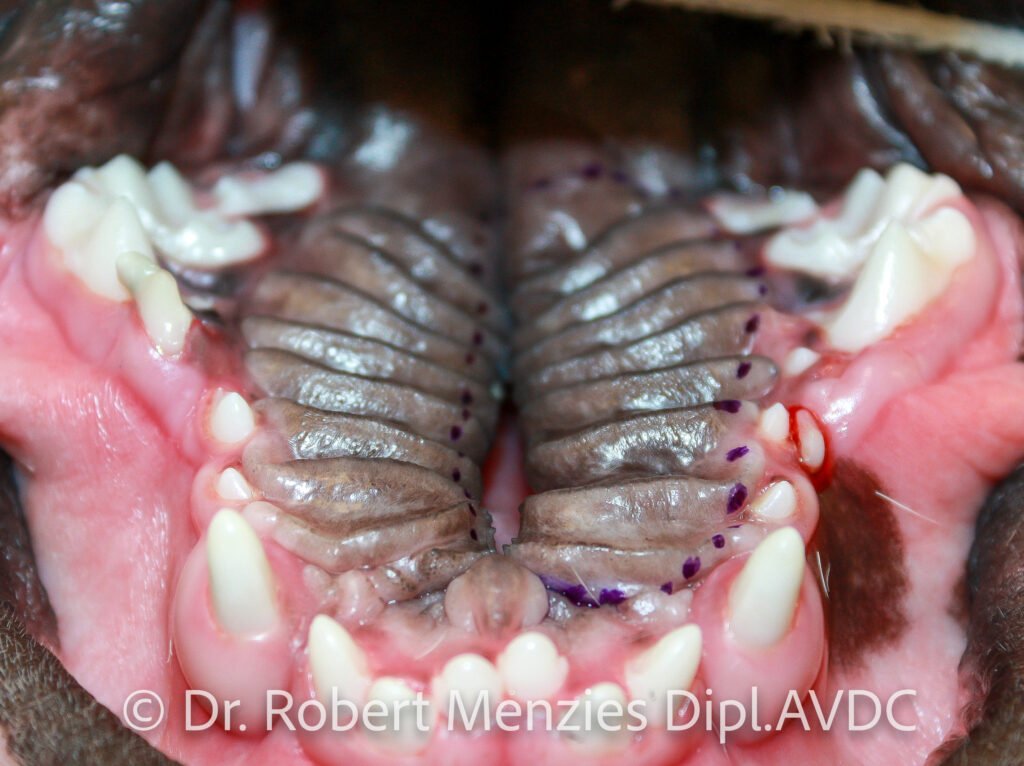
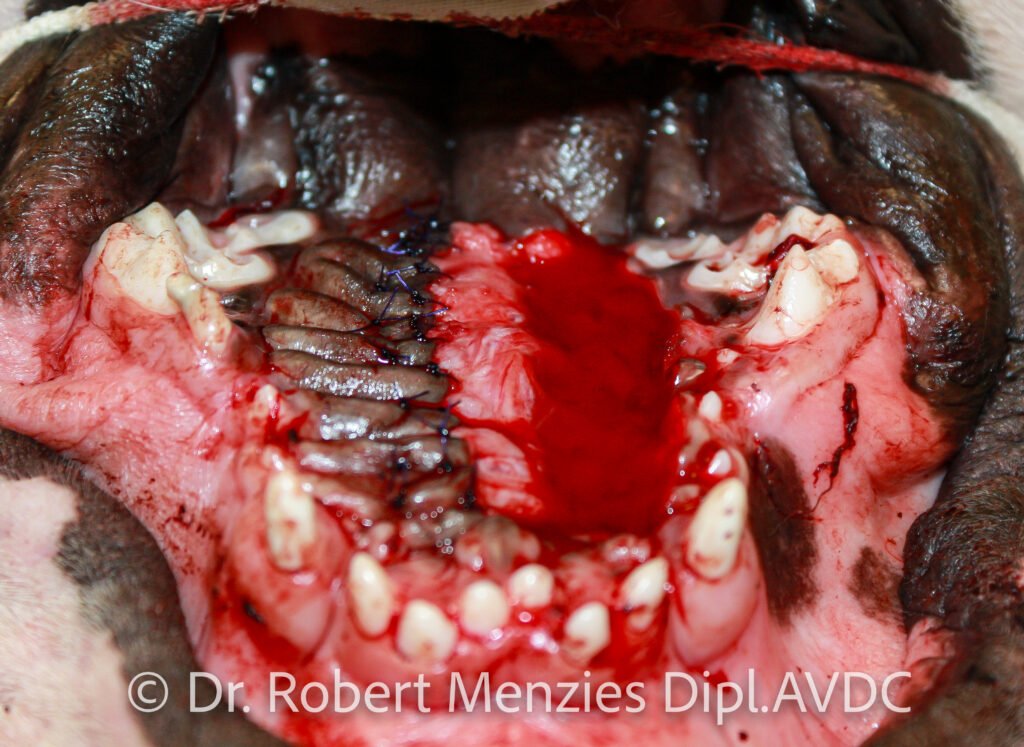
Oral Surgery
Interventions which involve the surgical disruption and manipulation of oral tissues and their supporting structures. Techniques may employ cold hard steel, electrosurgery and laser, amongst others. Commonly surgery is devoted to the extraction of teeth or their fragments, operculectomies for unerupted teeth, tumour and cyst resection, palatal defects and jaw fractures. Surgery may be employed in conjunction with other animal tooth treatment techniques such as a type III crown lengthening procedure, retrograde root canal therapy and periodontal surgeries.
Other surgeries involve the lips, tongue and salivary glands. Due to the jaws typically containing many tooth roots, traditional approaches to orthopaedic trauma such as plate and screw techniques may be problematic, and thus less invasive techniques involving interdental wiring and acrylic intraoral splinting may be indicated.
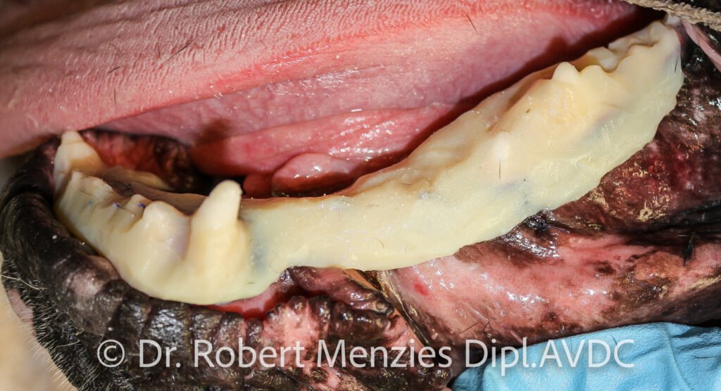
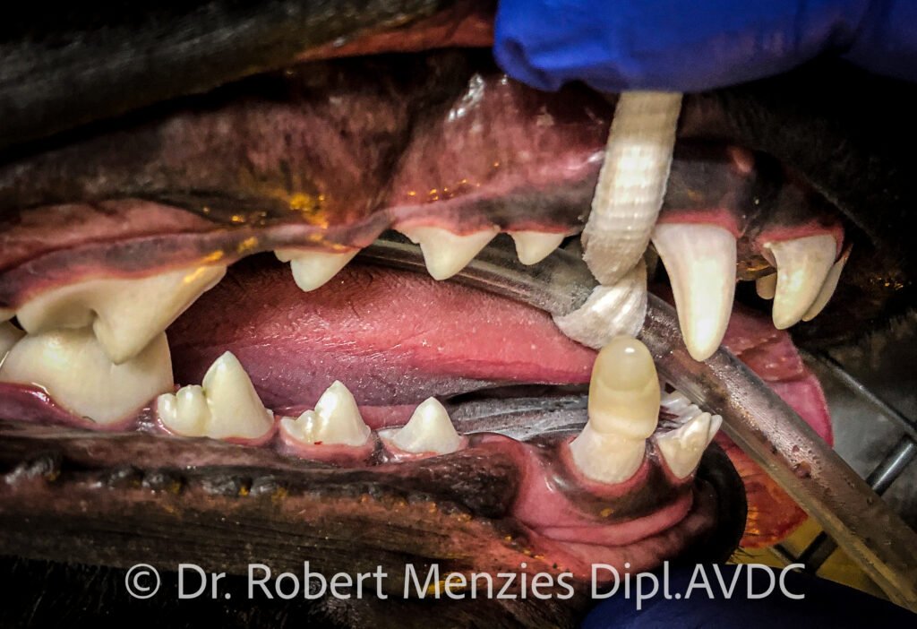
Extraction and debridement of hard palate
Palatal surgery
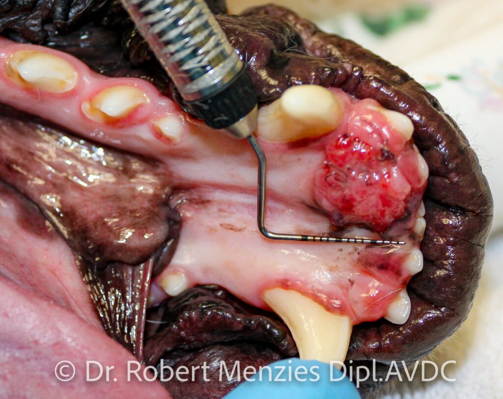
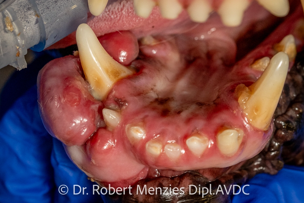
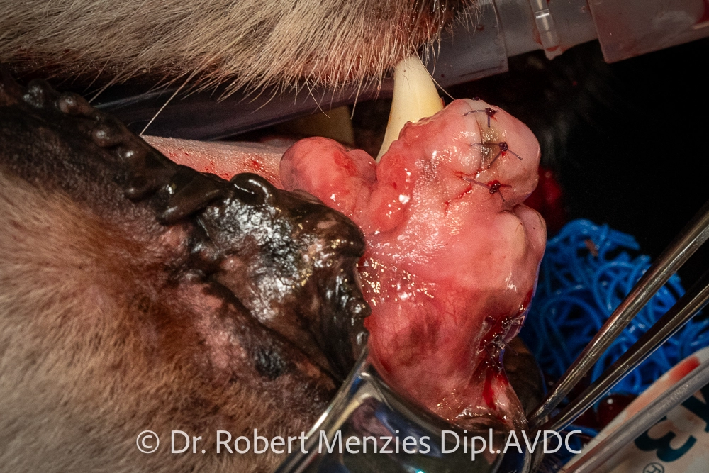
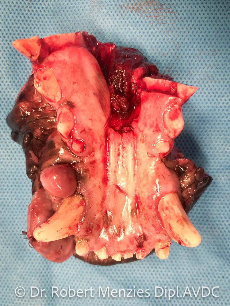
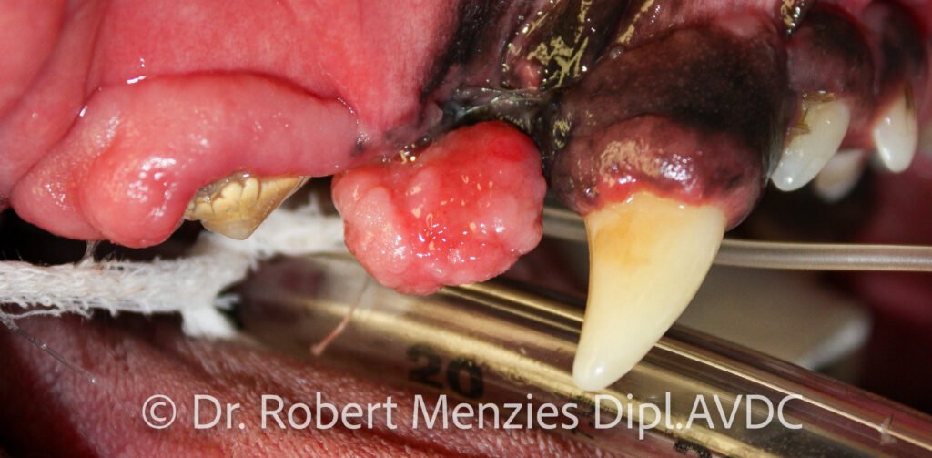
Oncologic surgery
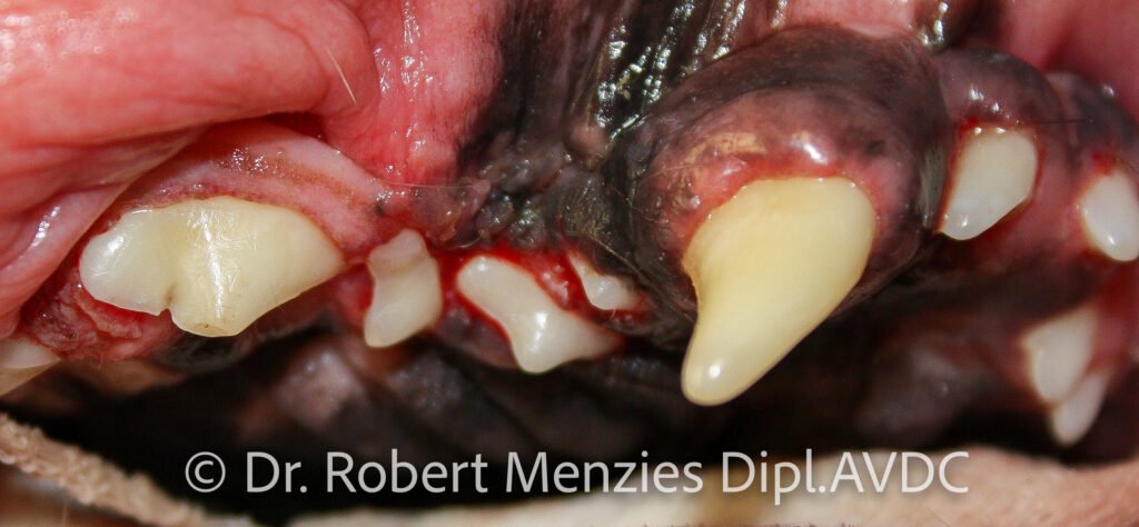
Jaw fracture repair

procedure.
Orthodontics
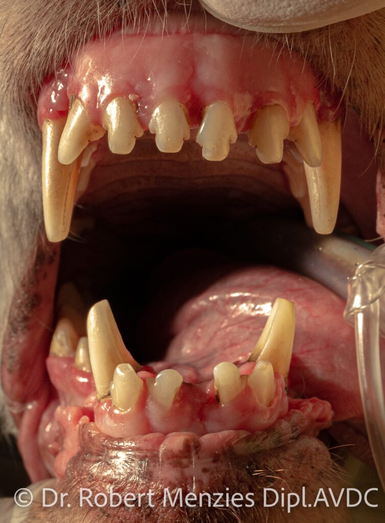
Orthodontics involve the diagnosis, management and treatment of dental and jaw abnormalities which lead to an abnormal bite. Classically, animal tooth treatment involves the movement of teeth either in the prevention or treatment of a traumatic or unfavourable malocclusion, however it also includes other interventions, such as, crown reduction and vital pulp therapy, and extraction. The malocclusion may be due to factors such as dental crowding, or upper and lower jaw growth discrepancies. Interceptive orthodontics are performed during development in an effort to prevent or reduce malocclusion in the mature animal. An orthodontic consult is advised as soon as a malocclusion is diagnosed to formulate the most appropriate plan. Persistent deciduous teeth (i.e. baby teeth which have not been shed as soon as the crown of the permanent successor is visualised) require careful removal as a matter of priority to limit the consequence of such a malocclusion. Less often teeth may be extracted in an effort to allow a jaw to grow to its potential without the inhibition of a dental interlock.
Passive orthodontics, such as “ball therapy”, temporary crown extensions or an acrylic bite plane for linguoverted canine teeth, are activated by the animal closing their mouth and thus they are in control of how much force they will tolerate on their teeth. With active orthodontics the degree of force applied to a tooth is dictated by the human, and may involve elastic bands, springs or screws. Depending on the type and complexity of the tooth movement required, one or the other type of appliance may be used, or a combination. Orthodontics comprise a journey and may not be suitable to all circumstances. Malocclusions which are of a developmental nature rather than acquired (e.g. due to trauma, surgery or a mass) very likely have a genetic component and subsequent breeding of the animal is strongly advised against.
Crown reduction

Temporary crown extensions
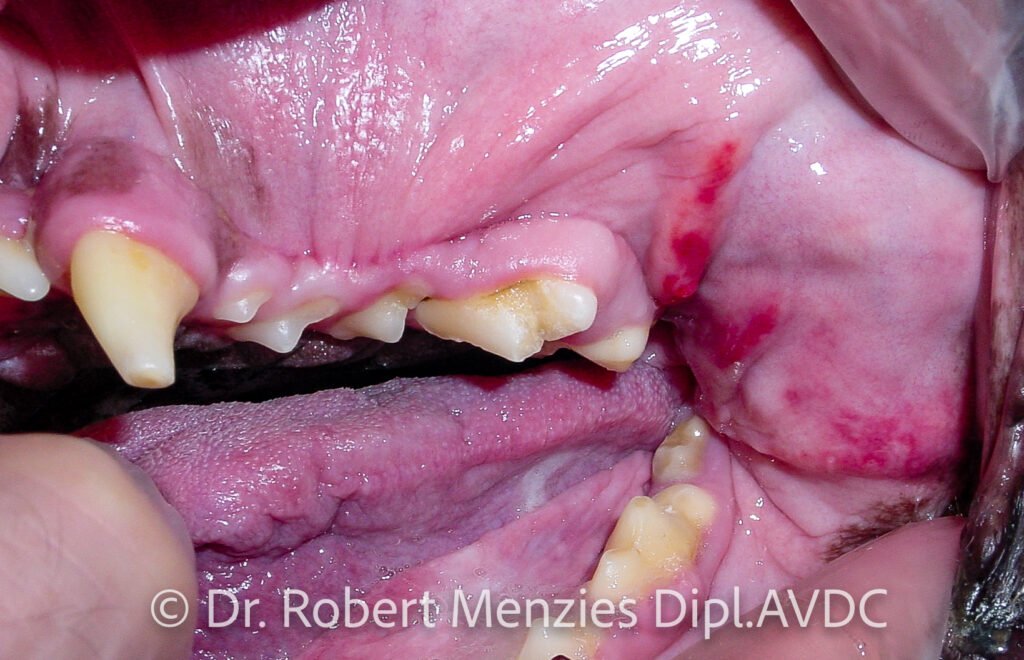
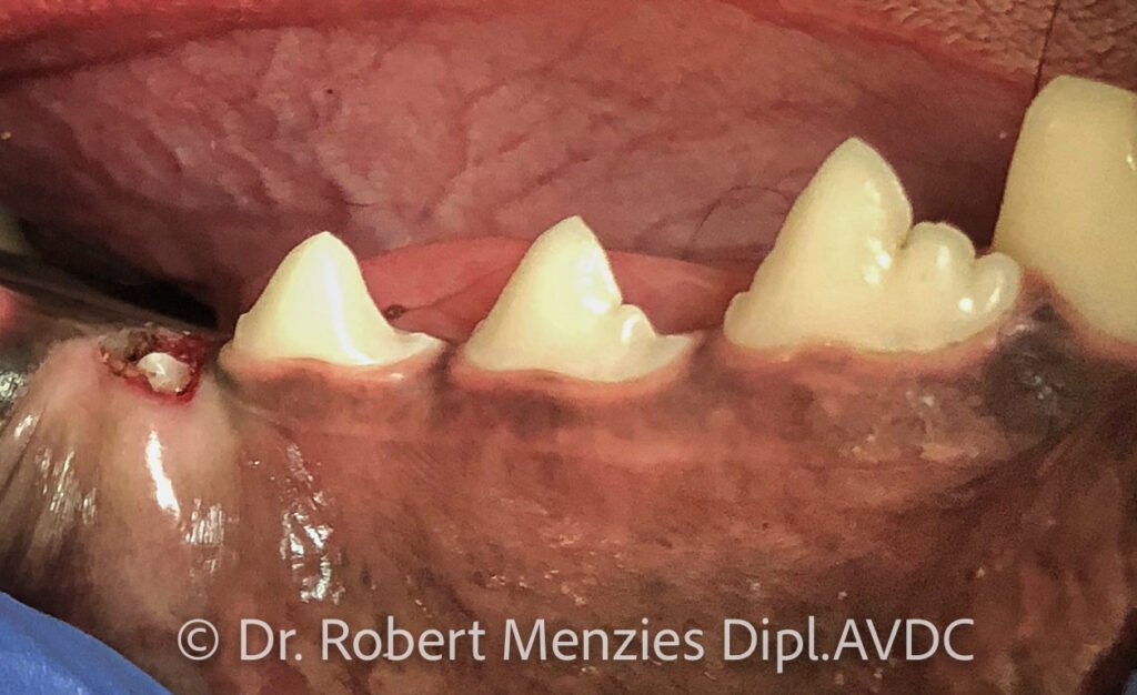
Periodontics
Periodontics are concerned with the health of the supporting structures of the tooth – gingiva, periodontal ligament, cementum and alveolar bone. It is influenced by the anatomy of the oral and supporting structures, immune system, diet, parafunction (such as chew toys, leisure and work activities) and home oral hygiene routines. An evaluation of periodontal health therefore requires an assessment of each of these areas. This is obtained through history taking, an awake examination and a detailed examination under general anaesthesia involving periodontal charting and further diagnostics where indicated such as dental radiography. Treatments for periodontal disease include a thorough dental cleaning, root planing, perioceuticals, and periodontal surgery, including guided-tissue regeneration. However, it is what happens at home on a day-to-day basis which has the biggest influence on periodontal health in the medium and long term.
Hence, engagement of the owners in their animal’s oral health is a crucial component of any successful treatment plan. The treatment of traumatic injuries is possible such as those due to dental malocclusion, laceration of the gingiva, and dental avulsion and luxation. Reimplantation of an avulsed tooth if you wish to save it is a dental emergency. Reading from the human literature, dental avulsions of permanent teeth should be replaced back in the alveolus (socket) as soon as possible (don’t replace avulsed deciduous teeth) and the tooth kept moist until this occurs with (in decreasing order of preference) milk, Hank’s Balanced Salt Solution (HBSS), saliva or saline – ideally within 15 minutes, okay within 1 hour, and a poor long-term prognosis for longer than an hour.
Gingivectomy – gingivoplasty
–
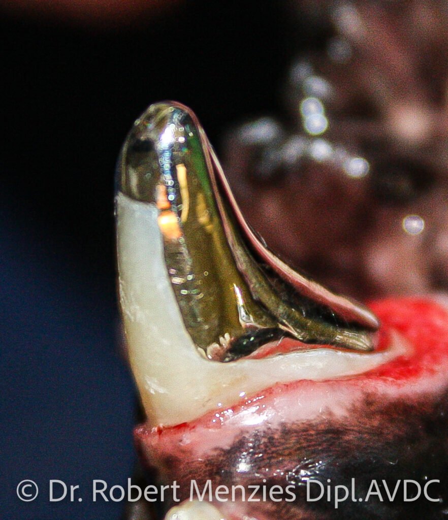
Prosthodontics
Prosthodontics are an animal tooth treatment referring to dental prostheses, and in the veterinary world is typically limited to prosthodontic crowns, and less so dental implants. Prosthetic crowns for the veterinary patient are usually made of metal due to their durability and are placed to help prevent further degradation of the crown. Full-metal crowns may be placed on non-vital teeth once their root canal therapies have been found to be successful. Partial crowns are preferred to be placed on vital (living) teeth due to the risk of irreversible pulpits occurring as a result of a full crown preparation. A three-quarter crown may be placed on the canines of a dog which has severe wear due to being a cage biter and who can’t be removed from the environment (for example, a police dog). Partial crowns could also provide protection to teeth with severe enamel defects. The placement of a crown takes a number of steps, typically over several weeks.
The steps occur under general anaesthesia. Initially, full-mouth dental impressions are obtained, a crown preparation is performed, followed by a detailed impression of the treated tooth. The dental impressions are sent to a dental laboratory and the metal crown is formed. Once the metal crown is received at the practice, the dog is anaesthetised again, the metal crown tried for fit, and it is cemented in place. The health of the tooth under the crown requires long-term monitoring. It is important to note that placing a prosthetic crown actually weakens the tooth due to the amount of tooth removed in performing the crown preparation, and thus the benefits to the animal of preventing ongoing wear versus the increased risk of breaking the tooth need to be appreciated with this animal tooth treatment.
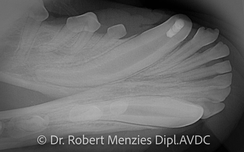
Prosthetic crowns in a police dog
left upper canine tooth.
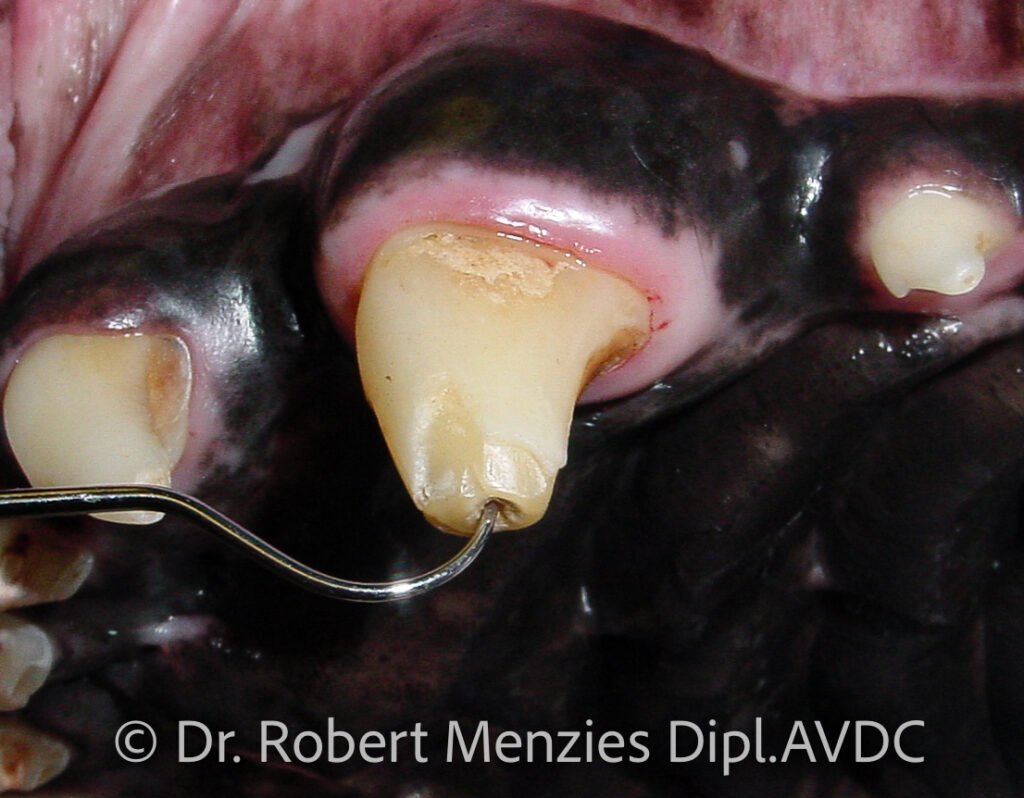
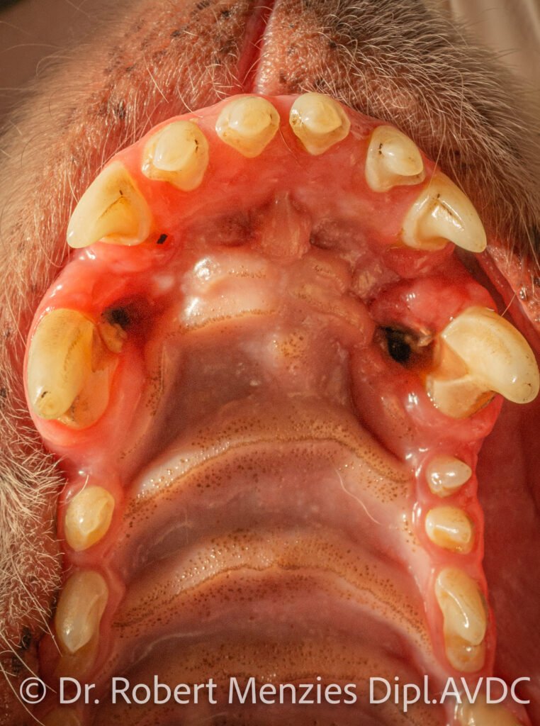
Restorations
Restorations may be placed to restore dental defects. In the veterinary world, tooth-coloured composite materials are commonly used. Dental defects may be developmental in nature, for example, amelogenesis imperfecta, however more typically they are acquired. Common examples of acquired dental defects include the inadvertent iatrogenic damage of enamel which may happen has a consequence of damaging the crown of an underlying unerupted tooth when extracting a deciduous tooth, trauma to the crown, and less commonly in animals, to repair the dental structures following carious lesions (dental ‘cavities’).
Restorations are also an animal tooth treatment used to restore an access site used to facilitate endodontic therapy, such as root canal therapy.
Composite restorations